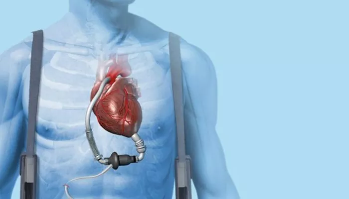
Heart failure (HF) is a complex clinical syndrome marked by the heart’s inability to pump blood effectively. Echocardiography (echo) plays a central role in diagnosing and monitoring HF. As a non-invasive, real-time imaging tool, it helps evaluate left ventricular function, chamber size, wall motion, and valvular integrity. Timely echo assessments guide clinical decisions, monitor disease progression, and assess therapy response. However, determining how often to repeat an echo depends on several variables. These include the HF type—whether reduced ejection fraction (HFrEF) or preserved ejection fraction (HFpEF), symptom changes, treatment modifications, or preoperative planning. This article explores evidence-based guidelines and expert consensus to establish appropriate intervals for echocardiographic monitoring in HF.
Why Echocardiograms Matter in Heart Failure
Echocardiograms provide vital structural and functional insights:
- Measure ejection fraction (EF)
- Assess valvular status
- Identify ventricular hypertrophy or dilation
- Evaluate diastolic function
- Detect pericardial effusion or thrombus
These findings influence medication adjustments, surgical decisions, and prognosis. Echo remains the standard initial and follow-up test due to its safety and accessibility.
General Guidelines for Echocardiogram Frequency
No universal timeline fits all HF patients. However, major guidelines, including those from the American College of Cardiology (ACC), American Heart Association (AHA), and European Society of Cardiology (ESC), offer broad recommendations:
- At initial diagnosis of heart failure
- After major changes in clinical status
- Following the introduction or escalation of disease-modifying therapies
- Every 6 to 12 months in stable HFrEF patients
- Every 1 to 2 years in stable HFpEF patients
These timelines shift based on patient condition, therapy response, or suspected complications.
Echocardiogram Timing by Heart Failure Type
Heart Failure with Reduced Ejection Fraction (HFrEF)
In HFrEF (EF ≤ 40%), EF measurements help monitor therapy response. ACE inhibitors, beta-blockers, ARNI, and aldosterone antagonists often improve EF.
Recommendations:
- At diagnosis: Baseline EF and chamber dimensions
- 3 to 6 months post-therapy change: Check for EF improvement
- Every 6-12 months if stable: Ongoing surveillance
- With new/worsening symptoms: Reassess immediately
Heart Failure with Preserved Ejection Fraction (HFpEF)
In HFpEF (EF ≥ 50%), echo tracks diastolic function and left atrial pressure indicators.
Recommendations:
- At diagnosis: Evaluate diastolic filling patterns
- With clinical deterioration: Repeat promptly
- Annually or biannually: For stable patients, especially elderly or hypertensive
Heart Failure with Mid-Range Ejection Fraction (HFmrEF)
EF between 41-49% represents a transition zone. Patients may progress toward either spectrum.
Recommendations:
- Baseline and follow-up: Echo every 6-12 months
- After interventions: Repeat 3-6 months post-treatment
Clinical Triggers for Repeat Echocardiograms
New or Worsening Symptoms
Dyspnea, fatigue, edema, or exercise intolerance may signal EF decline, valvular dysfunction, or new wall motion abnormalities.
Therapeutic Adjustments
Initiating ARNI or device therapy (ICD, CRT) requires reassessing EF to track reverse remodeling.
Pre-Surgical Evaluation
Patients undergoing non-cardiac surgery or valve replacement may need an updated echo within six months.
Post-Myocardial Infarction
HF post-MI demands EF surveillance to monitor for ventricular aneurysm, thrombus, or deterioration.
Device Therapy Considerations
Cardiac resynchronization therapy (CRT) success depends on improved synchrony and EF. Echo within 3-6 months post-implantation is standard.
Monitoring Valvular Heart Disease in HF
Valve dysfunction often coexists with HF. Echo tracks stenosis or regurgitation progression.
Frequency:
- Mild disease: Every 3-5 years
- Moderate disease: Every 1-2 years
- Severe disease: Annually or with symptom change
Echo in Advanced Heart Failure or Transplant Candidates
In stage D HF or transplant evaluation, serial echo evaluates pulmonary pressures, RV function, and LV end-diastolic dimension. This supports eligibility assessments and guides therapy.
Role of Point-of-Care Ultrasound (POCUS)
POCUS complements traditional echo by offering bedside assessment. In HF clinics or hospital settings, it enables rapid evaluation of:
- LV function
- Pericardial effusion
- Volume status via IVC measurement
While not a replacement for full echo, it reduces time to diagnosis and optimizes resource use.
When Not to Repeat Echocardiograms
Routine echo repetition without clinical change lacks value. Medicare and insurance guidelines discourage unnecessary testing.
Do not repeat if:
- No therapy change and patient is stable
- EF was recently measured (within past 6-12 months)
Judicious imaging avoids patient burden and healthcare costs.
Special Populations
HF in Pregnancy
Pregnant women with HF require tailored monitoring. Echo at baseline, mid-pregnancy, and postpartum ensures maternal-fetal safety.
Pediatric HF
In congenital or acquired pediatric HF, echo frequency depends on growth stages, surgical timing, and clinical status.
Geriatric HF
In older adults, consider comorbidities and frailty. Annual echo may suffice unless symptoms shift.
Limitations and Alternatives
Though echo is preferred, it has limitations in obese patients or those with poor acoustic windows.
Alternatives include:
- Cardiac MRI (for precise EF and fibrosis assessment)
- Nuclear imaging (for viability studies)
- CT angiography (for coronary or structural assessment)
These are used selectively when echo results are inconclusive.
Expert Consensus and Case-Based Adjustments
Echo frequency often depends on nuanced judgment. For example:
Case 1: A 58-year-old male with HFrEF started on ARNI. Echo at 6 months to assess remodeling.
Case 2: A 72-year-old woman with stable HFpEF and hypertension. Echo every 1.5-2 years unless symptoms worsen.
Case 3: A CRT recipient with non-ischemic cardiomyopathy. Echo 3 months post-implant to evaluate EF improvement.
Conclusion
The timing of echocardiograms in heart failure hinges on HF subtype, treatment status, symptoms, and overall stability.
Most patients benefit from echo at diagnosis, after clinical changes, and periodically for therapy monitoring. Overuse should be avoided in stable cases without therapeutic intervention. Individualized schedules—anchored in guidelines, but tailored by clinical context—ensure optimal care.
Related topics:


