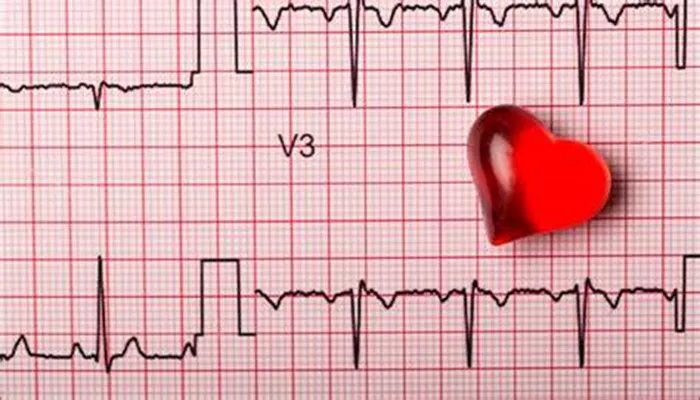
An electrocardiogram (EKG or ECG) is a common test to assess heart function. It records the electrical signals of the heart. Many patients ask if an EKG can detect blocked arteries. This question is important because blocked arteries can lead to serious heart problems, including heart attacks. In this article, we will explain what an EKG can and cannot do regarding blocked arteries. We will also describe other tests that help detect artery blockages more accurately.
What Is an EKG and How Does It Work?
An EKG measures the electrical activity generated by the heart as it beats. Electrodes placed on the skin detect tiny electrical changes. These changes create a graph showing the timing and strength of heartbeats. The EKG helps doctors see how the heart’s rhythm and electrical system function.
Basic Components of an EKG
- P wave: Shows atrial contraction
- QRS complex: Shows ventricular contraction
- T wave: Shows ventricular relaxation
Changes in these waves can suggest heart problems.
What Are Blocked Arteries?
Blocked arteries happen when the blood vessels supplying the heart become narrow or closed. This is mainly due to a buildup of fatty plaques called atherosclerosis. When arteries narrow, blood flow reduces, which can cause chest pain (angina), shortness of breath, or even a heart attack.
Common Causes of Blocked Arteries
- Atherosclerosis: Fat, cholesterol, and other substances form plaques inside artery walls
- Blood clots: Can suddenly block arteries, causing heart attacks
- Spasms: Temporary tightening of arteries
Can an EKG Detect Blocked Arteries?
In short, an EKG does not directly detect blocked arteries. It records electrical activity but does not show blood flow or artery narrowing. However, an EKG can sometimes suggest the effects of blocked arteries on the heart.
How EKG Shows Effects of Blockages
When arteries are blocked, parts of the heart may get less oxygen. This can cause changes in the heart muscle’s electrical activity. These changes may show on the EKG as:
ST segment changes: Elevation or depression may indicate ischemia (low oxygen).
T wave inversion: Shows abnormal repolarization due to poor blood flow.
Q waves: May indicate past heart attacks caused by blockages.
Limitations of EKG in Detecting Blocked Arteries
Normal EKG does not rule out blockages: Some patients with blocked arteries have normal EKGs, especially at rest.
Transient changes: Ischemic changes can be temporary and may not appear if the EKG is done when symptoms are absent.
Non-specific changes: EKG abnormalities can be caused by other heart conditions, not only blocked arteries.
When Does an EKG Help Identify Blocked Arteries?
During a Heart Attack (Myocardial Infarction)
During a heart attack, blood flow to the heart muscle stops due to blocked arteries. This causes clear changes on the EKG. Doctors use EKGs to diagnose heart attacks quickly. Specific patterns, such as ST elevation in certain leads, can show the location and severity of the blockage.
During Stress Testing
A resting EKG may miss blockages, but a stress EKG can help. The test records EKG during exercise or medication-induced stress. If blocked arteries limit blood flow during stress, ischemic changes may appear on the EKG. This helps identify patients who need further testing.
Other Tests That Detect Blocked Arteries More Accurately
Since EKG alone cannot reliably detect blockages, doctors use other tests for better diagnosis.
Stress Echocardiogram
This test combines ultrasound images of the heart with exercise or medication stress. It shows how the heart muscle moves and if parts do not get enough blood.
Nuclear Stress Test
This test uses a small amount of radioactive material to image blood flow in the heart during rest and stress. It highlights areas with poor blood supply due to blockages.
Coronary Angiography
This is the gold standard test. A catheter is inserted into arteries, and contrast dye is injected. X-ray images show the exact location and degree of blockages. It is invasive but very accurate.
CT Coronary Angiography
A non-invasive CT scan that uses contrast dye to visualize coronary arteries. It can detect narrowing or blockages without catheterization.
When Should You Get an EKG to Check for Blocked Arteries?
EKGs are often the first step when patients report chest pain, shortness of breath, or other symptoms of heart disease. It is quick, painless, and widely available. It helps rule out acute heart problems and guides further testing.
Symptoms Suggesting Blocked Arteries
- Chest pain or pressure
- Shortness of breath
- Fatigue with exertion
- Palpitations or dizziness
Routine Screening
In people without symptoms but with risk factors (e.g., diabetes, high cholesterol, smoking), an EKG may be part of a routine heart health check. However, a normal EKG does not exclude blockages.
Interpreting EKG Results in the Context of Blocked Arteries
Doctors interpret EKG results alongside patient history, physical exam, and other tests. EKG findings alone cannot confirm or exclude blocked arteries. They provide clues to the heart’s electrical health, which may be affected by blockages.
When EKG Is Normal
A normal EKG does not mean the arteries are clear. Many patients with significant blockages have no EKG changes at rest.
This is why further testing is needed if symptoms persist.
When EKG Shows Abnormalities
Abnormal EKG changes raise suspicion of ischemia or past heart attacks. Doctors will usually order stress tests or imaging to confirm artery status.
Conclusion
An EKG is a valuable, fast tool to assess heart electrical activity. It can show signs suggestive of blocked arteries during acute events like heart attacks or ischemia during stress. However, it cannot directly detect or rule out artery blockages. Other tests like stress echocardiography, nuclear imaging, and angiography are necessary for accurate diagnosis.
If you have symptoms or risk factors, speak with your healthcare provider about the best testing approach. Early detection and treatment of blocked arteries can save lives and improve quality of life.
Related topics:


