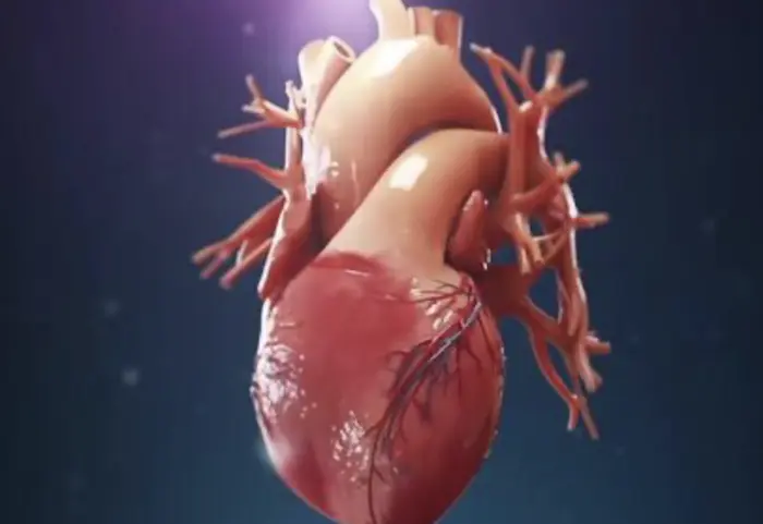
Myocardial infarction (MI), commonly called a heart attack, occurs when blood flow to the heart muscle is blocked. This causes damage or death to the heart tissue. One of the most dangerous complications of MI is ventricular fibrillation (VF), a rapid, irregular heart rhythm. VF can lead to sudden cardiac death if not treated immediately.
Understanding how MI causes VF is critical for timely intervention and improving patient outcomes.
What Is Ventricular Fibrillation?
Ventricular fibrillation is a severe cardiac arrhythmia. It happens when the ventricles of the heart quiver instead of contracting normally. This disorganized electrical activity prevents the heart from pumping blood effectively.
VF results in immediate loss of cardiac output and, if untreated, causes death within minutes.
Characteristics of Ventricular Fibrillation
- Rapid, erratic electrical impulses in the ventricles
- No coordinated ventricular contraction
- Loss of effective heartbeat and blood circulation
- Causes sudden collapse and unconsciousness
How Myocardial Infarction Triggers Ventricular Fibrillation
Myocardial infarction damages heart muscle and disrupts its electrical system. Several mechanisms link MI to the development of ventricular fibrillation.
Ischemia-Induced Electrical Instability
During MI, blocked coronary arteries reduce oxygen supply to heart muscle cells. This ischemia causes metabolic changes that alter the electrical properties of the cells.
Ischemic cells lose their ability to maintain normal ion gradients.
This leads to abnormal depolarization and repolarization of cardiac cells.
The electrical instability can trigger premature ventricular contractions.
These abnormal beats can start a reentrant circuit causing VF.
Reentry Circuits and Arrhythmogenesis
MI creates areas of damaged and healthy tissue close together. This difference in tissue conductivity sets up reentry circuits.
Reentry is a phenomenon where electrical impulses loop repeatedly in the heart tissue.
This causes continuous stimulation of the ventricles without organized contraction.
Reentry circuits are a common cause of VF after MI.
Automaticity Changes in Damaged Cells
Ischemic and injured cells may develop abnormal automaticity. This means they can spontaneously generate electrical impulses.
- These ectopic impulses can disrupt the heart’s normal rhythm
- In combination with reentry, this leads to chaotic ventricular activity
Electrolyte and pH Imbalance
Ischemia causes shifts in potassium, calcium, and hydrogen ions in heart cells.
- High extracellular potassium shortens the refractory period
- Acidosis from lactic acid accumulation further impairs cell function
- These changes favor the initiation of VF by enhancing electrical instability
Structural Changes Post-Infarction
MI leads to scar formation and fibrosis in the ventricular myocardium.
- Scar tissue does not conduct electricity properly
- This heterogeneity in conduction slows and fragments impulses
- It promotes the persistence of reentry circuits and VF
The Timeline of Ventricular Fibrillation after Myocardial Infarction
VF can occur at different times following MI. Understanding the timing helps guide treatment and prevention.
Early VF (Within Minutes to Hours)
Most cases of VF after MI happen early. This is when ischemia and acute injury are most severe.
- Electrical instability peaks in the first hour
- Prompt reperfusion therapy can reduce this risk
Late VF (Days After MI)
VF can also occur days after MI during the healing phase.
Fibrosis and scar tissue develop, creating abnormal conduction paths.
Patients with large infarcts or poor heart function are at higher risk.
Risk Factors for VF in Patients with Myocardial Infarction
Not every patient with MI develops VF. Several factors increase the risk:
Large Infarct Size
More extensive myocardial damage means greater electrical disruption.
Reduced Left Ventricular Function
Weakened heart muscle is more prone to arrhythmias.
Electrolyte Imbalances
Low potassium or magnesium levels increase VF risk.
Previous History of Arrhythmias
Patients with prior ventricular arrhythmias have a higher chance of VF.
Delayed or No Reperfusion Therapy
Early reopening of blocked arteries lowers VF risk significantly.
Diagnosis and Monitoring of Ventricular Fibrillation after MI
VF is diagnosed using an electrocardiogram (ECG). It shows a chaotic, irregular waveform without distinct QRS complexes.
Continuous ECG monitoring is essential in MI patients, especially during the first hours after symptom onset.
ECG Features of Ventricular Fibrillation
- No organized QRS complexes
- Irregular, rapid electrical activity
- Absence of P waves and normal rhythm
Other Diagnostic Tools
Electrophysiological studies and imaging can help assess arrhythmia risk after MI.
Treatment and Prevention of Ventricular Fibrillation in MI
Immediate treatment of VF is critical to save lives. Long-term prevention focuses on managing MI and reducing arrhythmia triggers.
Emergency Management of VF
- Immediate defibrillation is the most effective treatment
- Advanced cardiac life support protocols include CPR and medications
Reperfusion Therapy
Opening blocked arteries early with thrombolysis or angioplasty reduces VF incidence.
Medications
Beta-blockers lower the risk of ventricular arrhythmias.
Antiarrhythmic drugs may be used in selected cases.
Electrolyte correction is important.
Implantable Cardioverter-Defibrillator (ICD)
In patients at high risk of recurrent VF, ICD implantation is recommended. It detects and terminates dangerous arrhythmias automatically.
Lifestyle Modifications
Healthy diet, regular exercise, quitting smoking, and controlling blood pressure help prevent MI and related VF.
Conclusion
Myocardial infarction creates multiple electrical and structural changes in the heart. These changes promote ventricular fibrillation, a life-threatening arrhythmia. Early recognition, treatment, and prevention strategies are vital to reduce morbidity and mortality.
Advances in reperfusion therapy and arrhythmia management have improved survival rates. Still, continuous research and awareness are essential to better understand and combat VF after MI.
Related topics:


