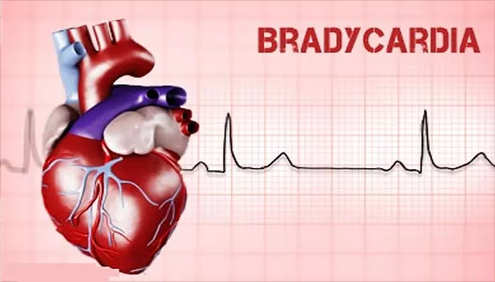
Primary bradycardia refers to an abnormally slow heart rate originating from intrinsic abnormalities in the heart’s conduction system, rather than external influences like medications or systemic illness. A healthy adult heart typically beats between 60 and 100 times per minute. When it beats below 60 beats per minute due to intrinsic cardiac causes, the condition is classified as primary bradycardia. This article explores the etiologies, pathophysiology, and clinical implications of this condition from a cardiology expert’s perspective.
Definition and Classification
Bradycardia is categorized by etiology into primary and secondary types. Primary bradycardia stems from structural or functional impairment within the cardiac conduction system itself. It may involve the sinoatrial (SA) node, atrioventricular (AV) node, or specialized conduction fibers. This is distinct from secondary bradycardia, which results from metabolic, pharmacologic, or systemic causes.
Physiology of Cardiac Conduction
To understand primary bradycardia, one must first understand the normal cardiac conduction system. The SA node, located in the right atrium, initiates the electrical impulse. This impulse travels through atrial myocardium to the AV node, then to the bundle of His and Purkinje fibers, stimulating ventricular contraction.
When any of these structures are compromised, the rate and rhythm of cardiac impulses slow, leading to bradycardia. In primary bradycardia, such impairment is typically due to degenerative or congenital defects.
Etiologies of Primary Bradycardia
Sinus Node Dysfunction (SND)
One of the most common causes of primary bradycardia is sinus node dysfunction. This condition includes several patterns:
- Sinus bradycardia
- Sinus arrest
- Sinoatrial exit block
- Chronotropic incompetence
Degenerative fibrosis of the SA node or surrounding atrial tissue is frequently observed in older adults. Fibrotic replacement leads to decreased pacemaker activity and conduction delay. In younger individuals, inherited channelopathies affecting the HCN4 gene may cause congenital SND.
Atrioventricular (AV) Conduction Disease
Another primary cause involves conduction abnormalities at the AV node or His-Purkinje system:
- First-degree AV block
- Mobitz type I and II second-degree AV block
- Third-degree (complete) AV block
Primary AV block often arises due to idiopathic fibrosis of the conduction system (Lenègre’s or Lev’s disease). These degenerative conditions affect older individuals, often without underlying heart disease.
Congenital Causes
In neonates and infants, congenital bradycardia is frequently observed in:
Congenital complete AV block
Congenital SND
Complete AV block in newborns may result from maternal autoimmune disease, such as lupus, where maternal anti-Ro/SSA antibodies cross the placenta and damage fetal cardiac conduction tissue. Genetic syndromes, like long QT syndrome or familial forms of SND, also contribute to congenital bradyarrhythmias.
Idiopathic Fibrosis of the Conduction System
Primary idiopathic fibrosis without associated cardiac pathology remains a major cause of conduction delay. Known as Lenègre disease (isolated fibrosis in younger patients) and Lev disease (age-related calcific degeneration), these conditions disrupt impulse transmission, especially in the His-Purkinje system.
Infiltrative and Degenerative Cardiac Disorders
Although some infiltrative diseases also involve extracardiac tissues, when they primarily affect the conduction system, they are considered primary bradycardia causes:
- Amyloidosis
- Sarcoidosis
- Hemochromatosis
These conditions deposit abnormal substances in cardiac tissue, replacing normal conduction fibers and impairing function.
Ion Channelopathies
Inherited mutations in genes encoding cardiac ion channels—sodium, calcium, and potassium—may lead to bradycardia by impairing impulse generation or propagation:
- HCN4 mutation (associated with sinus bradycardia)
- SCN5A mutation (may cause overlap of conduction disease and Brugada syndrome)
These rare but significant causes can manifest as isolated bradycardia or mixed arrhythmic phenotypes.
Post-Surgical and Post-Ablation Changes
Patients undergoing cardiac surgeries or catheter-based ablation may develop primary bradycardia if nodal tissue is inadvertently damaged. Though this may also be considered iatrogenic, the localized and direct injury to the conduction system classifies it as a primary defect in some contexts.
Age-Related Changes and Bradycardia
Ageing is a prominent risk factor for primary bradycardia. With age, fibrotic replacement of pacemaker tissue becomes common. This natural degeneration:
- Reduces SA node automaticity
- Delays AV conduction
- Increases risk of escape rhythms and syncope
Although often asymptomatic, elderly patients may develop symptomatic bradycardia necessitating pacemaker therapy.
Genetic Syndromes Linked to Primary Bradycardia
Several hereditary syndromes predispose to bradycardia through gene mutations:
- Familial Sick Sinus Syndrome
- Progressive Cardiac Conduction Disease (PCCD)
- Andersen-Tawil Syndrome
These disorders frequently present in childhood or early adulthood and may accompany structural cardiac anomalies or sudden cardiac death.
Pathophysiological Mechanisms
Multiple mechanisms lead to primary bradycardia:
- Reduced automaticity of SA node
- Blocked or delayed conduction through AV node or His-Purkinje system
- Abnormal impulse propagation due to fibrotic, infiltrative, or calcific changes
These effects disrupt regular cardiac pacing, reduce heart rate, and impair adequate cardiac output in some individuals.
Clinical Presentation
Symptoms vary based on heart rate and compensatory mechanisms:
- Fatigue
- Dizziness
- Syncope or near-syncope
- Palpitations
- Exertional intolerance
Severe cases with third-degree AV block or sinus arrest may result in Stokes-Adams attacks, requiring urgent intervention.
Diagnostic Evaluation
Electrocardiogram (ECG)
ECG remains the primary diagnostic tool. Key findings include:
- Prolonged PR interval
- Dropped beats
- Junctional or idioventricular escape rhythmy
Holter and Event Monitoring
Ambulatory monitoring detects intermittent or paroxysmal bradycardia. It is useful in evaluating unexplained syncope or episodic symptoms.
Electrophysiologic Studies
Invasive studies help define conduction intervals and localize dysfunction in complex or ambiguous cases. They aid in identifying candidates for pacemaker implantation.
Imaging and Laboratory Testing
Although imaging is not diagnostic for bradycardia, echocardiography may reveal structural heart disease. Laboratory tests may help rule out secondary causes (e.g., thyroid dysfunction, electrolyte imbalance), solidifying the diagnosis of primary bradycardia.
Management Strategies
Observation
Asymptomatic bradycardia, especially in athletes or during sleep, may not require intervention. Close monitoring suffices in mild cases.
Pacemaker Therapy
Symptomatic or hemodynamically significant bradycardia often requires permanent pacemaker implantation. Indications include:
- Sinus node dysfunction with syncope
- Advanced AV block
- Chronotropic incompetence
Device selection depends on rhythm, age, comorbidities, and lifestyle.
Genetic Counseling
In hereditary bradycardia, family screening and genetic counseling are crucial. Identifying carriers enables early intervention and risk stratification.
Prognosis
Prognosis depends on the cause and treatment. Idiopathic or age-related primary bradycardia generally responds well to pacemaker therapy. Congenital and genetic causes carry a variable prognosis based on associated arrhythmias and structural heart disease.
Conclusion
Primary bradycardia arises from intrinsic defects in the cardiac conduction system. Causes range from age-related fibrosis and genetic syndromes to congenital abnormalities and idiopathic degeneration. Diagnosis relies on thorough clinical evaluation and ECG monitoring. Treatment, often involving pacemaker therapy, can markedly improve quality of life.
Cardiologists play a pivotal role in recognizing and managing this complex condition, ensuring both accurate diagnosis and appropriate intervention.
Related topics:


